")
")
")
")
")
 |
|
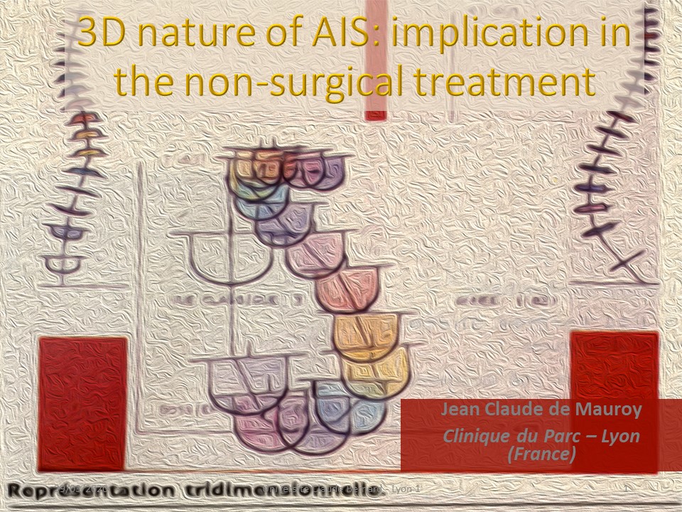 |
Scoliosis is a three-dimensional deformity of which all biomechanical characteristics are used for bracing, and physiotherapy. We will focus especially on the coupled movements that are the basis of the digital cast and can be used for the realization of all braces. You see in the background the first 3D representation of scoliosis done by Jean Dubousset and Henry Graf 50 years ago. The Da Vinci view was born. |
 |
Some fundamental rules of biomechanics are essential to understand scoliosis in its 3 dimensions. |
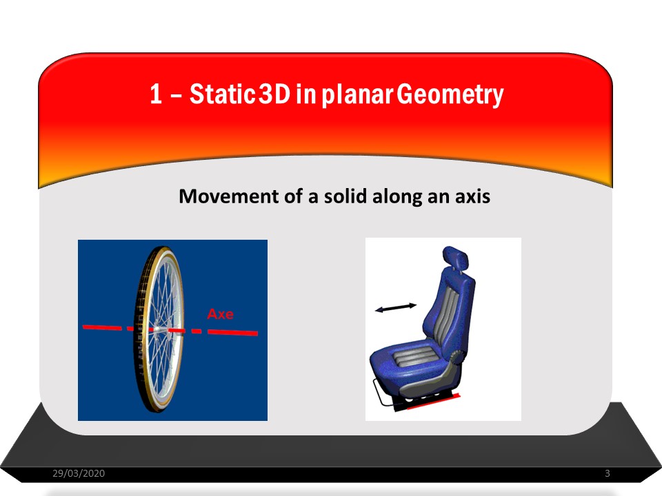 |
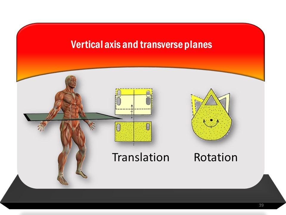
Around an axis, only two movements are possible: rotation and translation. |
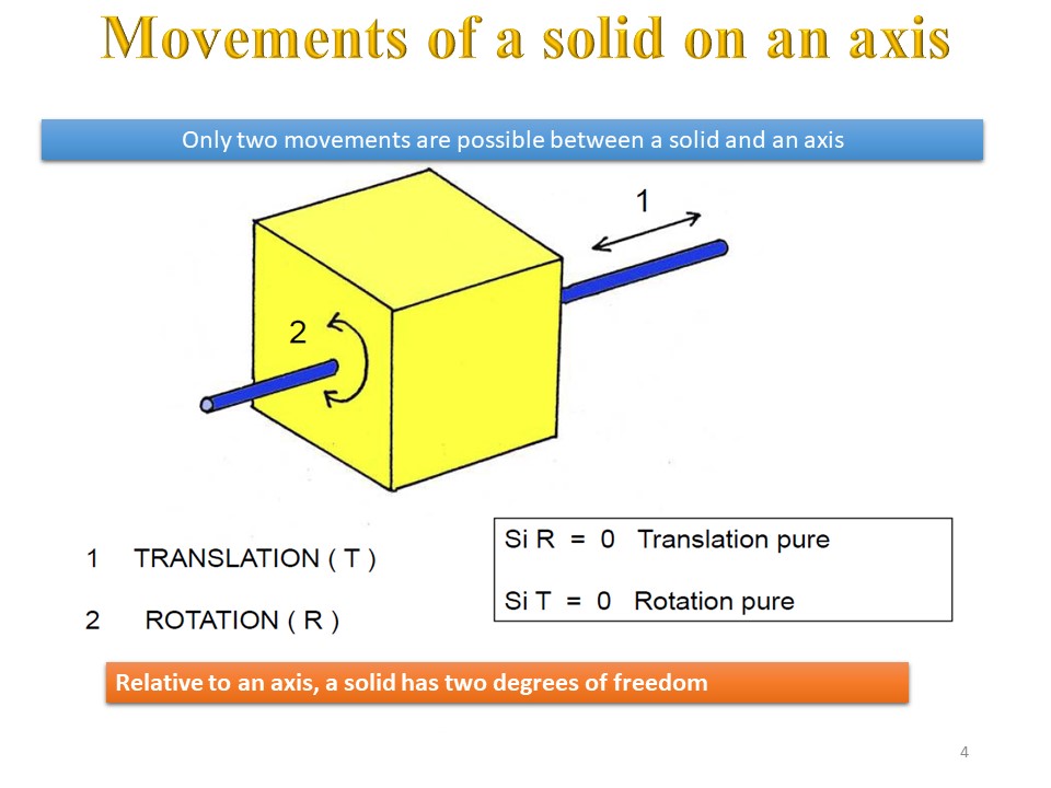 |
With respect to an axis, a solid therefore has two degrees of freedom. |
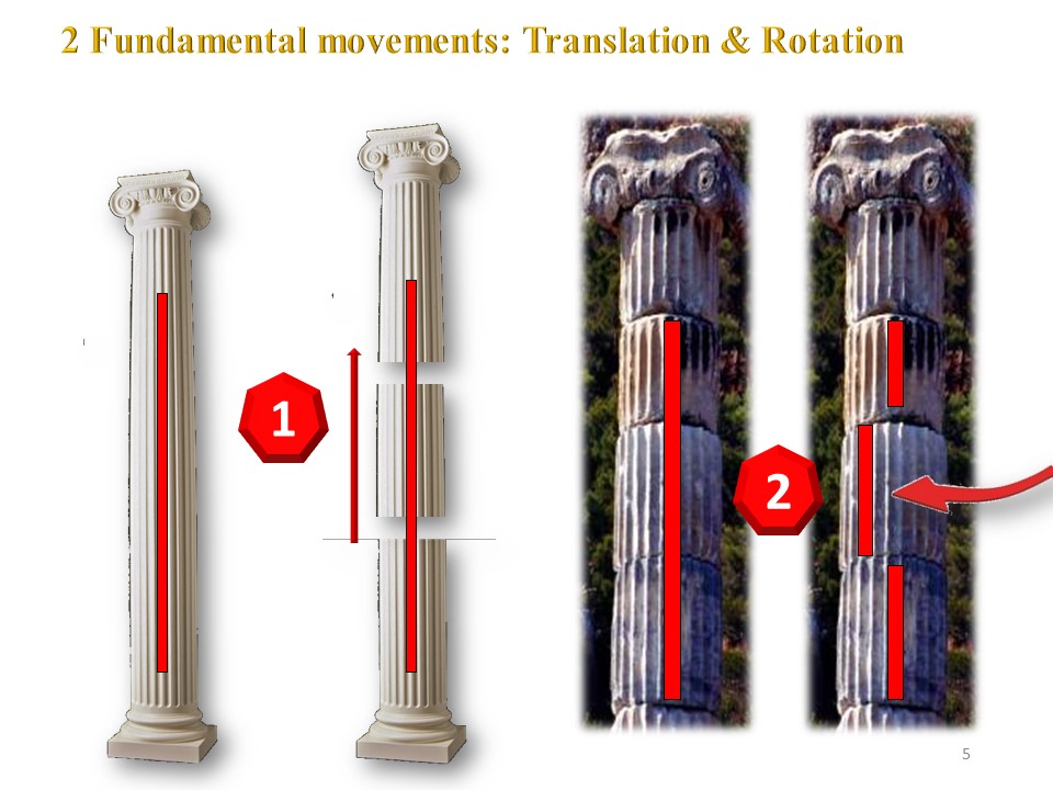 |
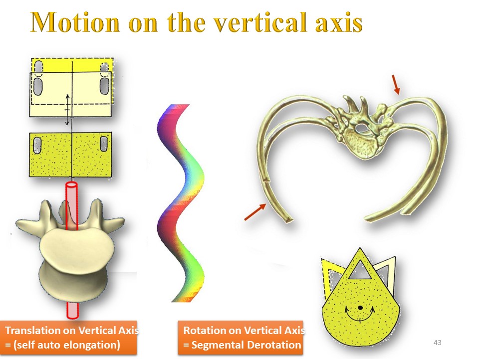
Translation and rotation for example along the vertical axis. |
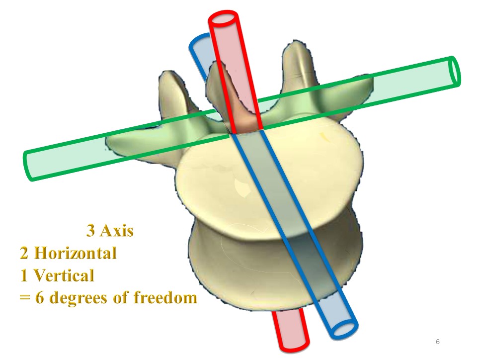 |
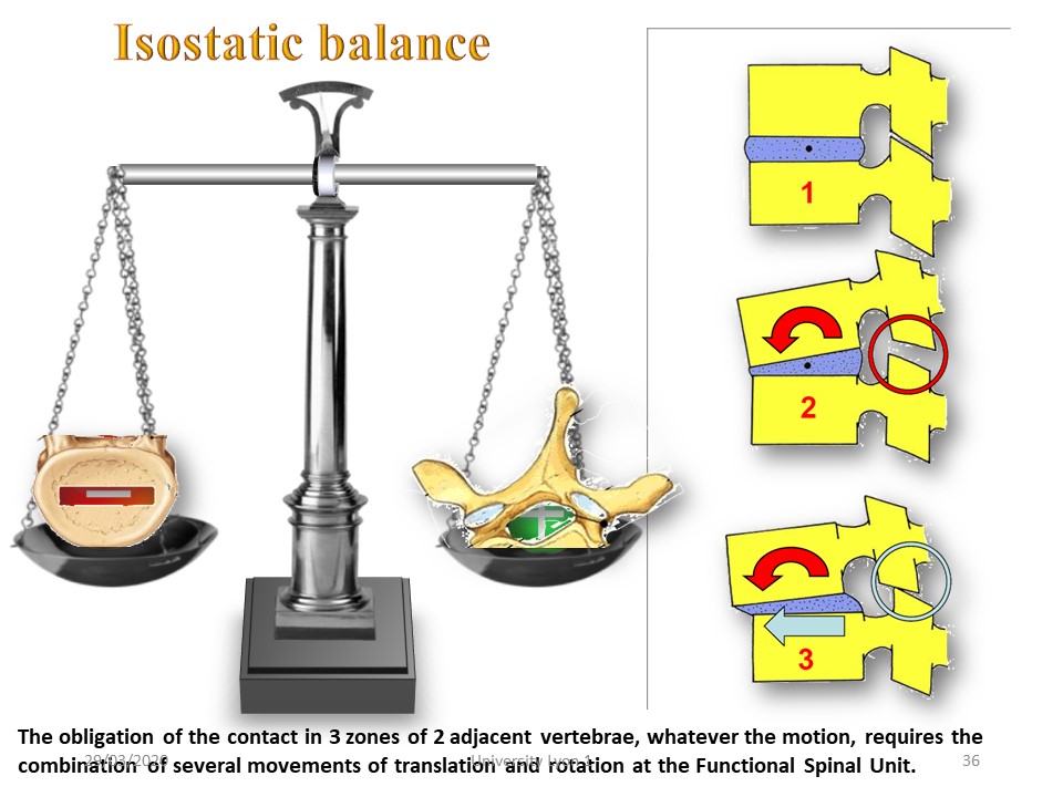
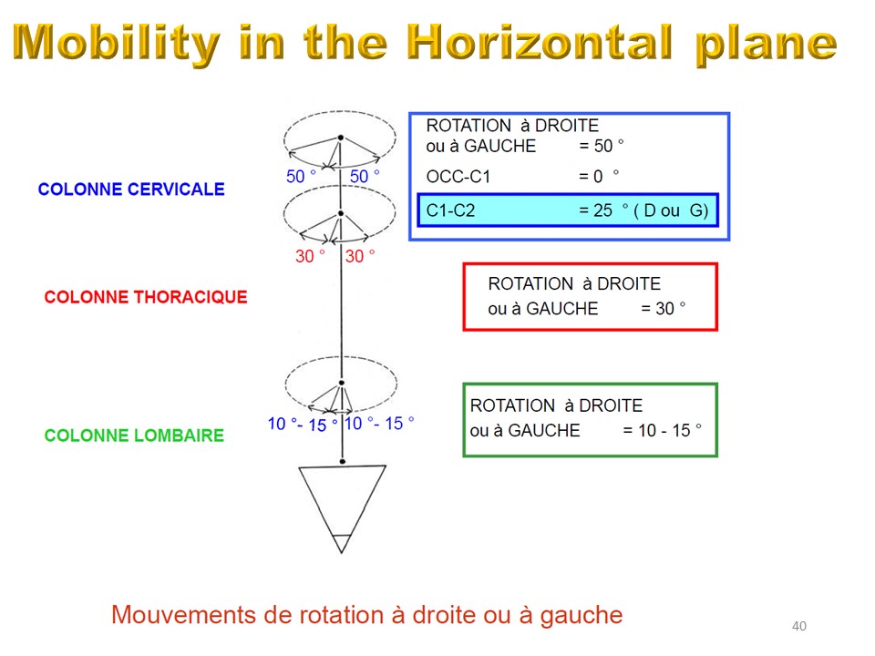
The movement between two vertebrae takes place along 3 axes: 2 horizontal; transverse (joining the transverse processes) and sagittal (in the axis of the spinous process) and 1 vertical (in the axis of the spinal cord), The intervertebral movement therefore has 6 degrees of freedom at each of the 17 thoracic and lumbar levels . |
 |
Successively the function plan of the spine and the 3 anatomical reference plans will be described. |
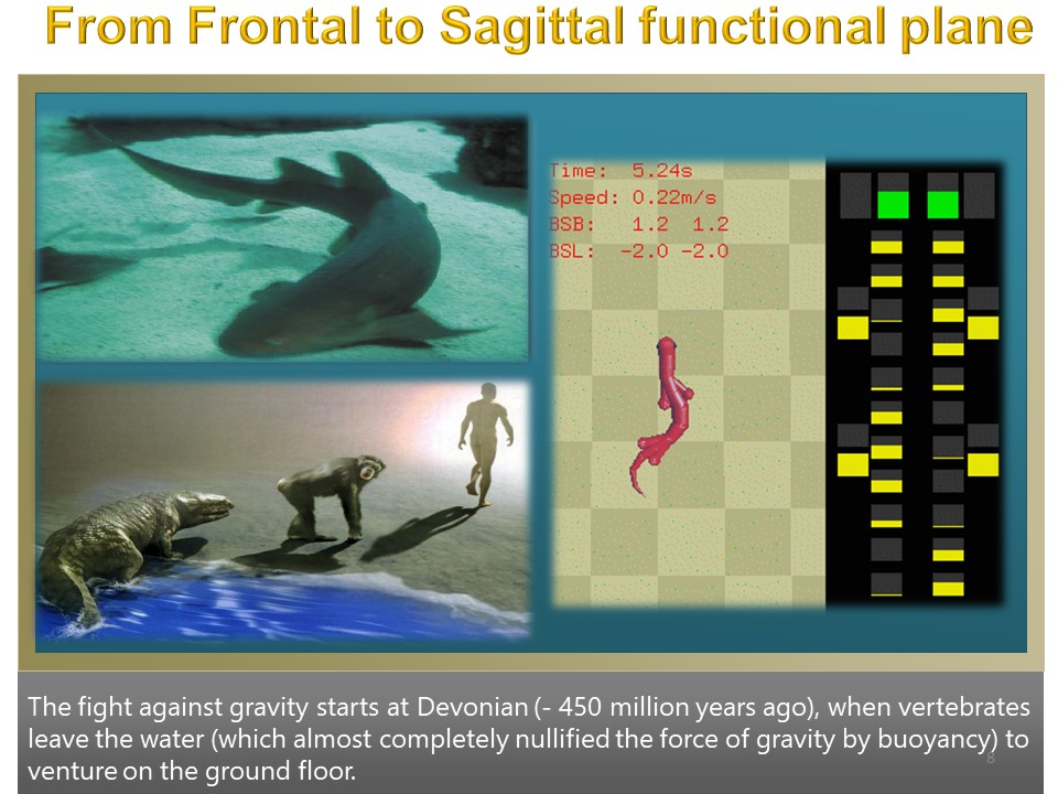 |
The initial function plane of vertebrates is the frontal plane. The passage from the aquatic environment to the terrestrial environment and the bipedalism modify the function plane, which becomes sagittal. |
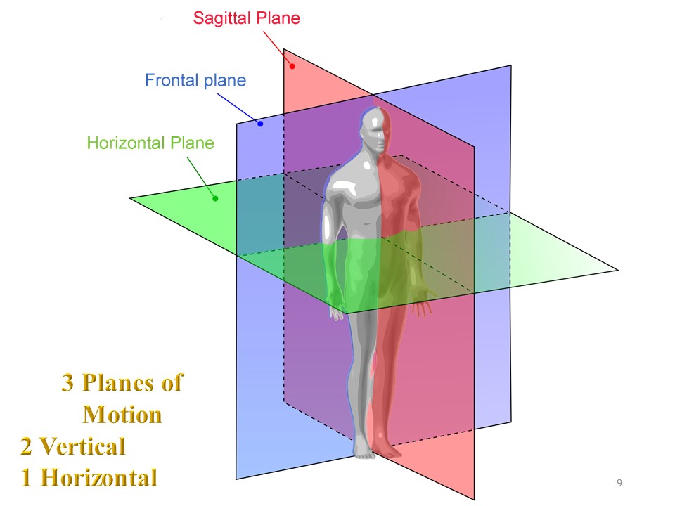 |
The 3 anatomical reference planes are: the frontal plane, the sagittal plane and the horizontal plane (or transverse plane). Although the movement takes place in space, by convention, the mobility of the spine is described in these 3 anatomical reference planes. |
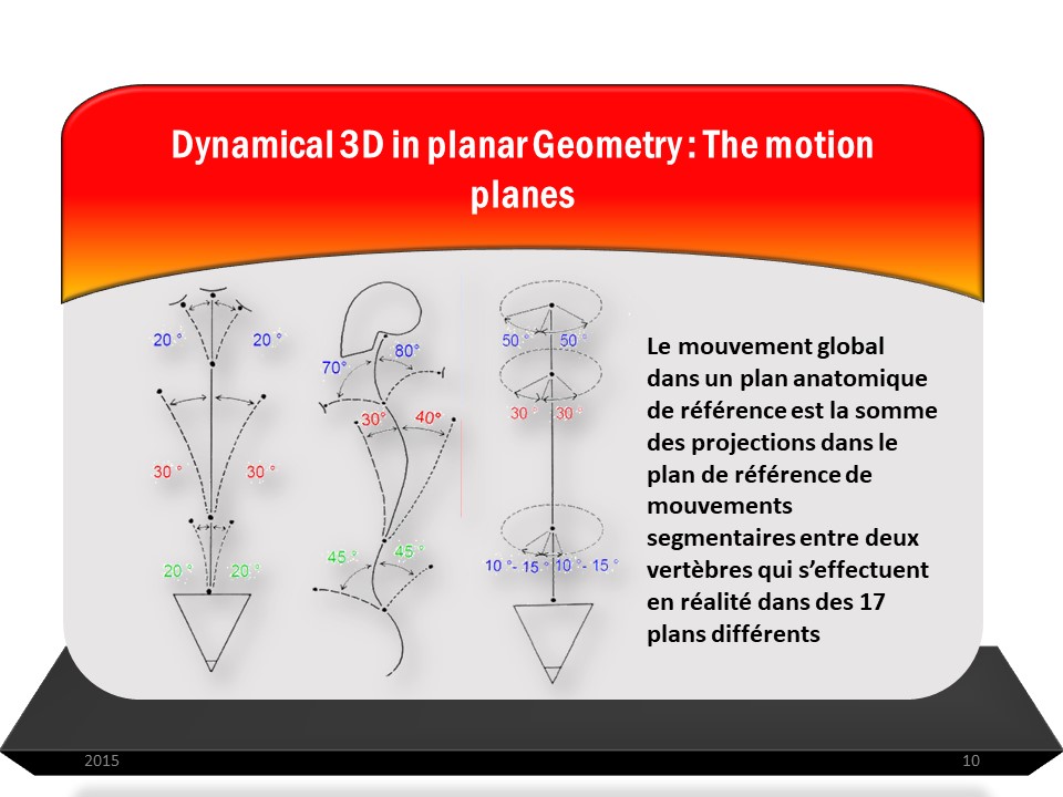 |
The overall movement in a reference anatomical plane is the sum of the projections in the reference plane of segmental movements between two vertebrae which are actually carried out in 17 different planes. |
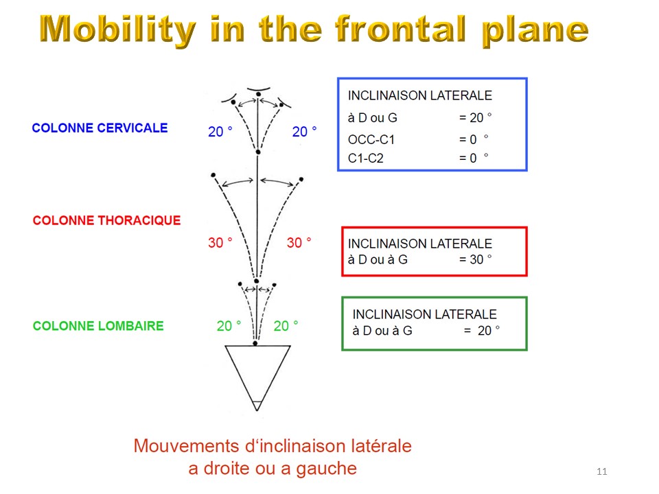 |
The movement in the frontal plane is the sum of all the intervertebral movements, at the lumbar, thoracic and cervical level |
 |
|
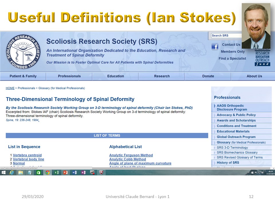 |
The 3D terminology has been defined by Ian Stokes and can be found on the Scoliosis Research Scoliosis website. |
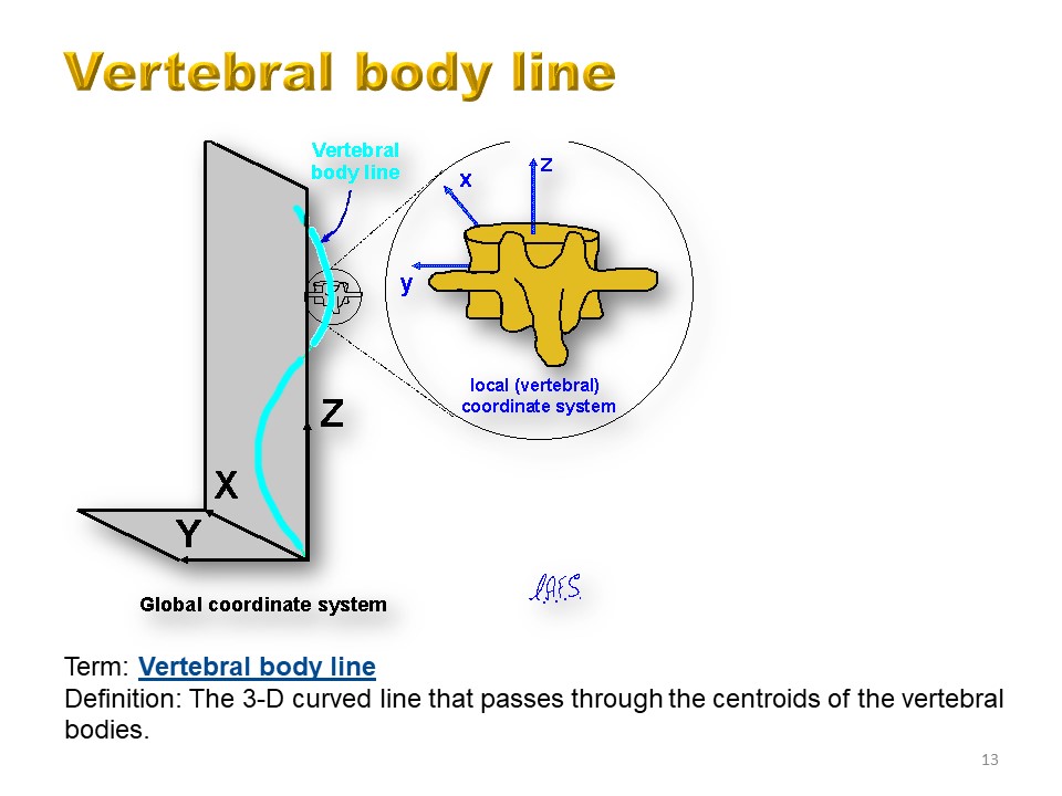 |
The vertebral body line describes the form of the spine in the global coordinate system. The vertebrae lie on this line. Each vertebra defines its own coordinate system depending on the its orientation. |
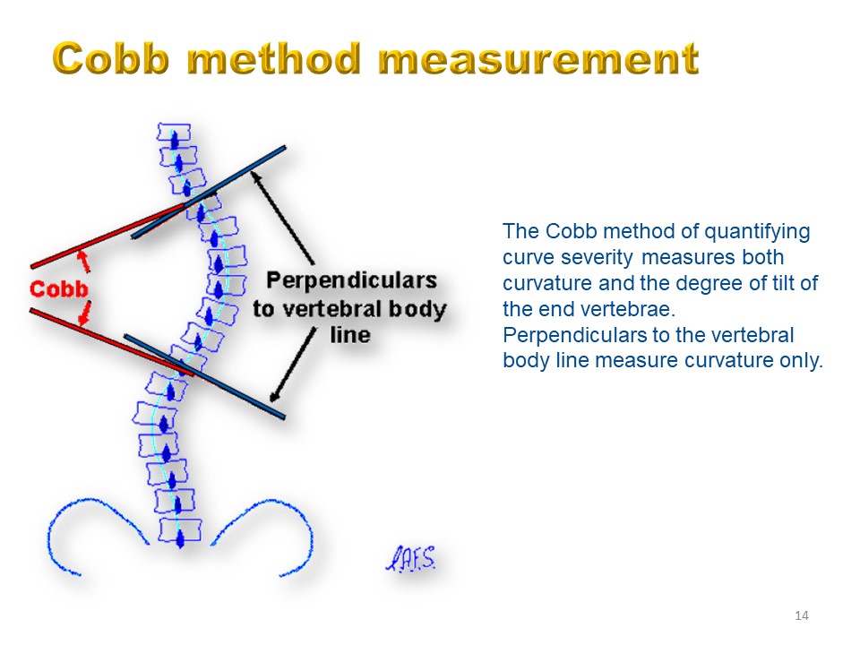 |
The Cobb method of quantifying curve severity measures both curvature and the degree of tilt of the end vertebrae. Perpendiculars to the vertebral body line measure curvature only. |
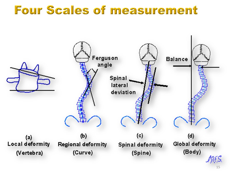 |
4 measurement scales can be used. The segmental level measures the deformation of the vertebral body, The regional level measures one of the 3 thoracic, lumbar and cervical regions. The spine level measures the entire spine. The overall level includes the pelvis and the head. |
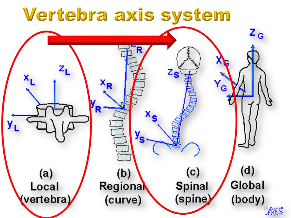 |
The hierarchy of four coordinate systems defining spinal geometry: (a) Local coordinates based on a vertebra. (b) Regional, curve-based coordinates defined by the end vertebrae of a curve or other spinal region. (c) Spinal coordinates defined by the Z axis passing through the centers of the most caudal and cephalad vertebrae of the entire spine. (d) Global, whole body-based coordinate system, with its origin at the base of the spine (S1), and the Z axis vertical (gravity line). |
|
The hierarchy of four coordinate systems defining spinal geometry: (a) Local coordinates based on a vertebra. (b) Regional, curve-based coordinates defined by the end vertebrae of a curve or other spinal region. (c) Spinal coordinates defined by the Z axis passing through the centers of the most caudal and cephalad vertebrae of the entire spine. (d) Global, whole body-based coordinate system, with its origin at the base of the spine (S1), and the Z axis vertical (gravity line). |
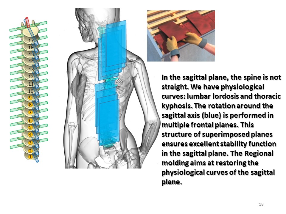 |
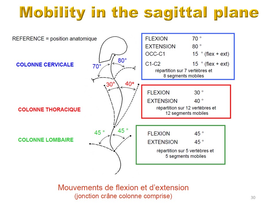
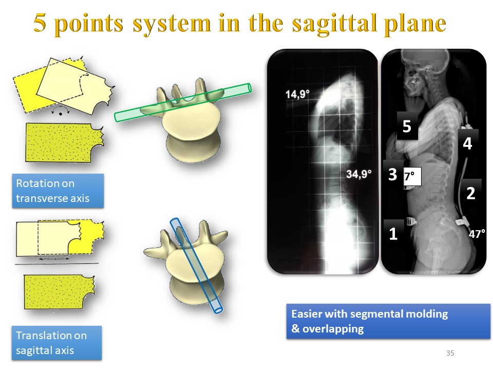
In the sagittal plane, apart from the flat back, the spine is not straight. We have physiological curvatures: lumbar lordosis and thoracic kyphosis. The rotation around the sagittal axes (blue) takes place in multiple planes This structure of superimposed planes ensures excellent stability in the sagittal function plane. The regional moulding aims to restore the curvatures of the sagittal plane. |
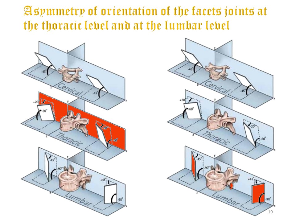 |
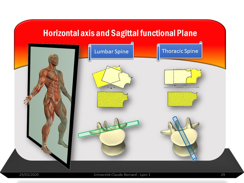
It is the orientation of the facet joints that will determine mobility in the frontal plane. At the thoracic level the facet joints are located in a frontal plane, while at the lumbar level they are located in a more sagittal plane. |
 |
|
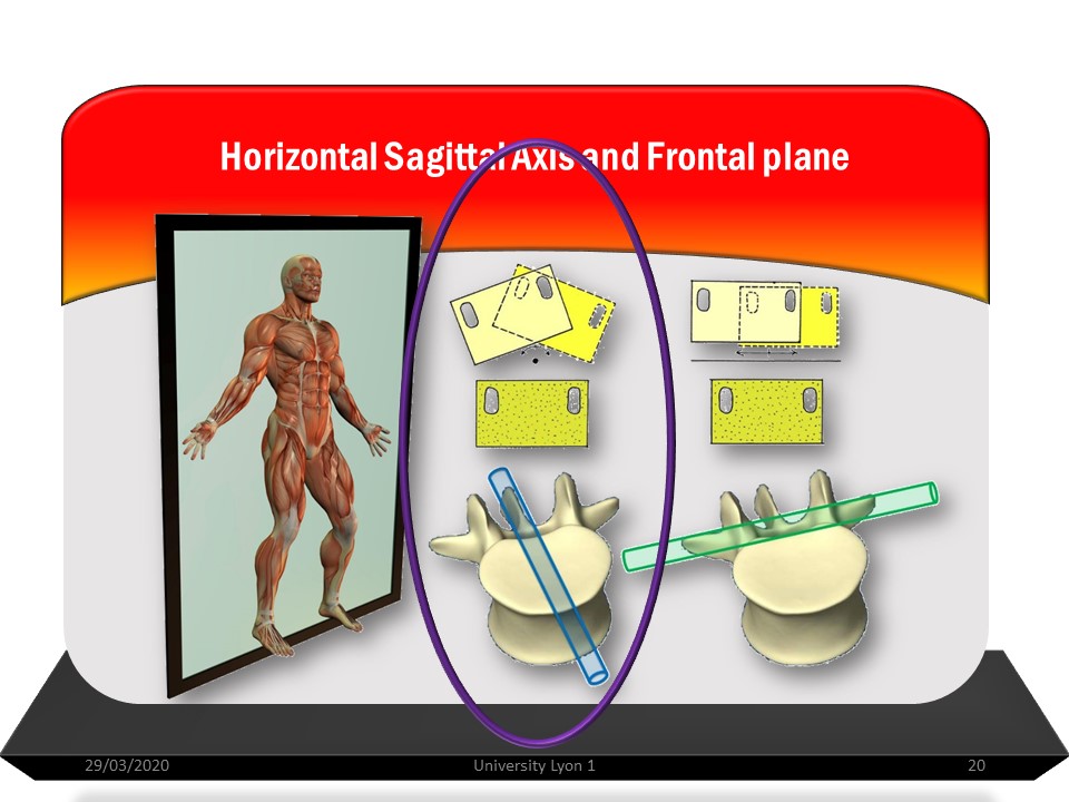 |
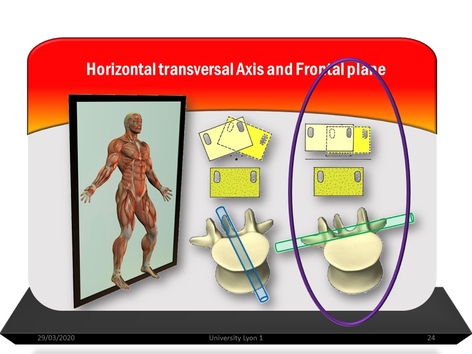
The movement in the frontal plane is obtained by rotation on the sagittal axis and / or translation along the transverse axis according to the orientation of the facet joints. If the column were straight, there would be only one frontal plane, considering the deviations in the frontal plane and in the sagittal plane, all the intervertebral movements are carried out in different frontal planes. |
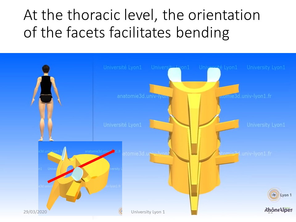 |
At the thoracic level, the frontal orientation of the facet joints favours rotation on the sagittal axis, that is to say the bending, |
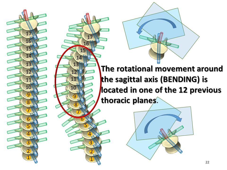 |
The rotational movement around the sagittal axis is in one of the 17 planes determined by the curvatures in the sagittal plane. |
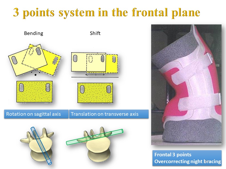 |
Some braces work according to this principle of bending induced by a 3-point system. |
 |
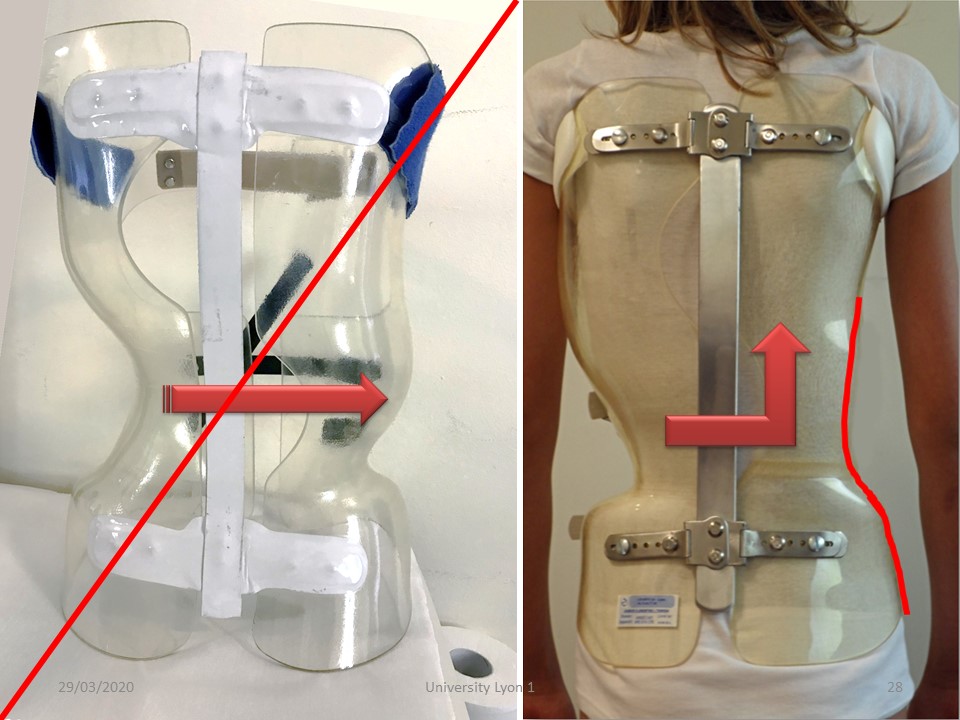
At the lumbar level, the sagittal orientation of the facet joints favours movements in the sagittal plane. Mobility in the frontal plane then takes place by translation along the transverse axis. |
 |
At the lumbar level, this translational movement along the transverse axis will be favoured by the thickness of the intervertebral disc and the absence of costo-vertebral joint. |
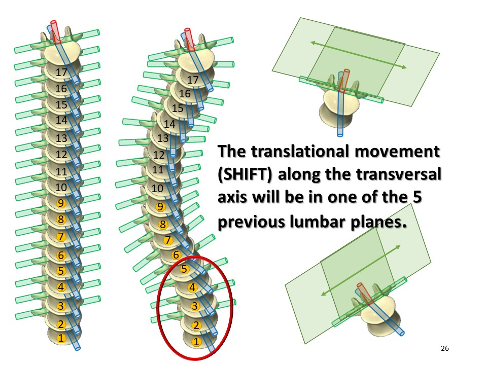 |
As before, this movement takes place in several planes due to lordosis |
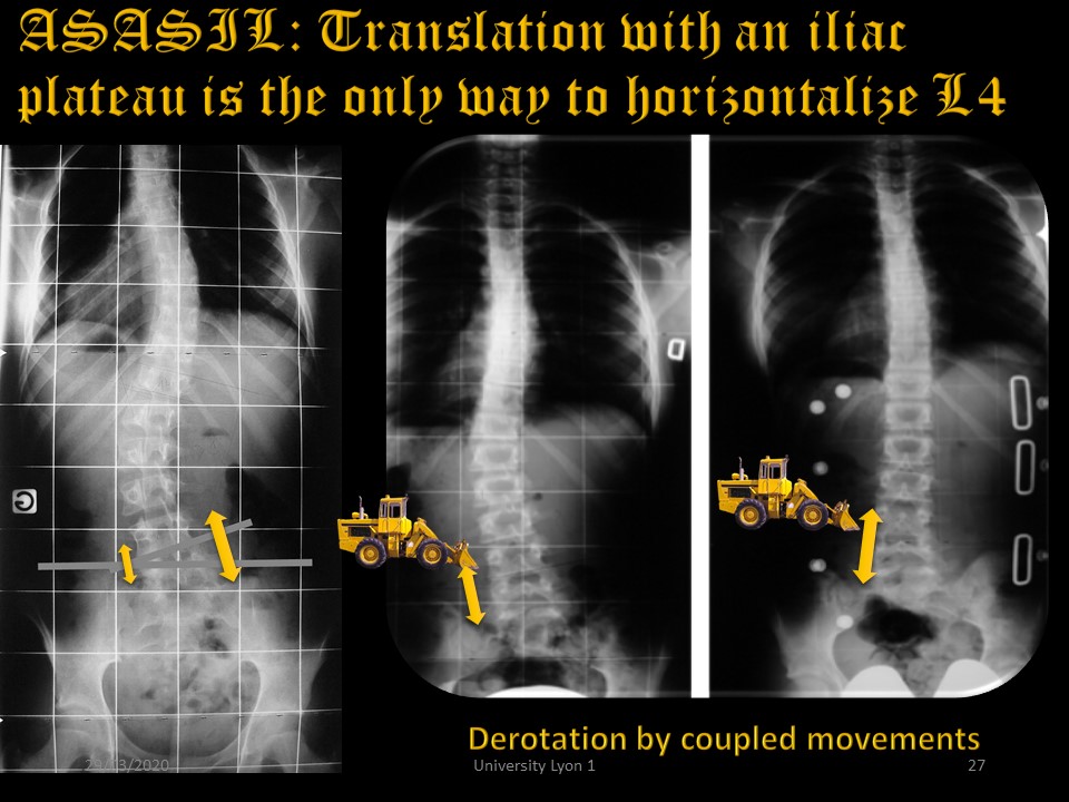 |
A second anatomical element can limit the lumbar shift. It is the Asymmetric Structural Anomaly of the Ilio-lumbar junction which was described by du Peloux 45 years ago. It is characterized by a tilt of L4 with shortening of the convex ilio-transversal ligament. The only way to stretch this ligament is to push the vertebral bodies of L3 and L4 toward the midline, with a shear at the iliac crest. |
 |
On the right, the shear at the level of the convex iliac plate is clearly visible. The expansion in the concavity is useless, on the contrary it is necessary to create a concave wall to propel the forces of frontal translation upwards (push up effect). In case of double curvature, the concave wall facilitates the corrective flexion of the thoracic convexity. |
 |
|
 |
In the sagittal plane the fundamental movements are also carried out at the level of the horizontal axes with inversion of the rotations and translations with respect to the frontal plane. Movement in the sagittal plane is the combination of rotation around the horizontal transverse axis and translation along the horizontal sagittal axis. |
 |
The movement in the sagittal plane is the sum of all the intervertebral movements at the lumbar, thoracic and cervical level |
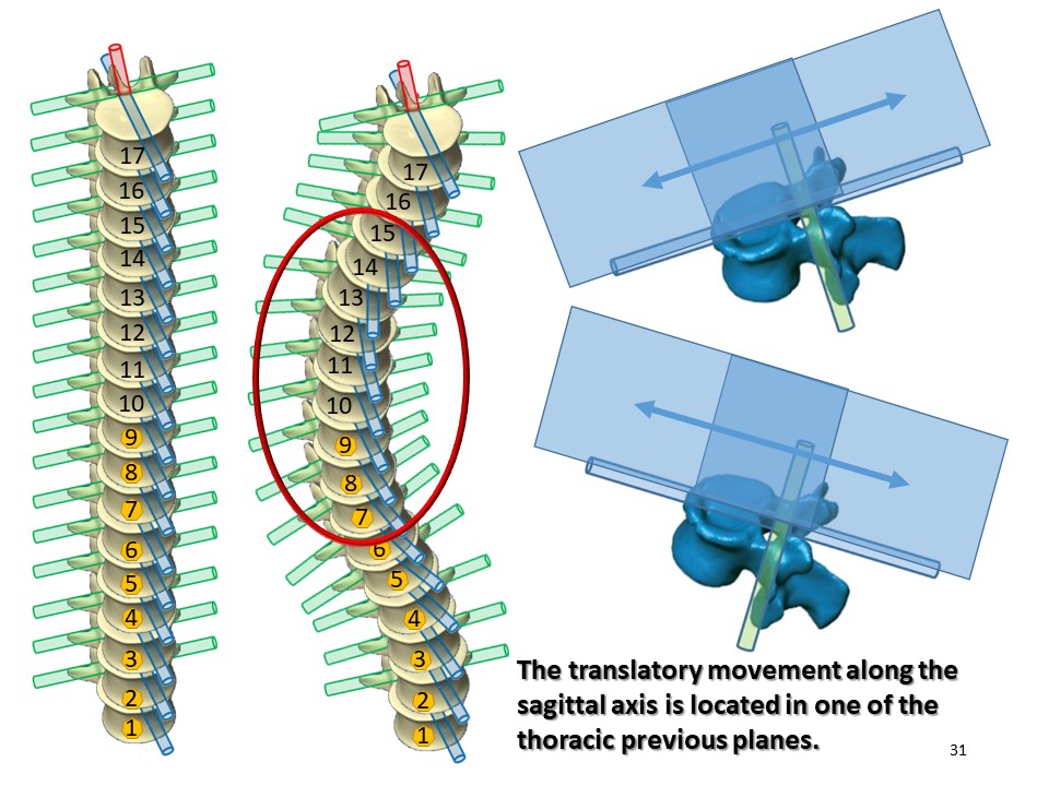 |
The translational movement along the sagittal axis is in as many planes as there are vertebrae included in the scoliotic curvature, essentially at the thoracic level taking into account the orientation of the facet joints. |
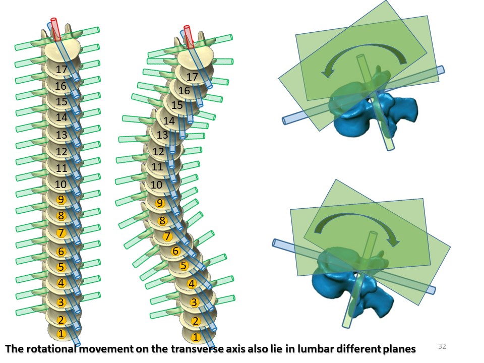 |
It is the same for the rotational movement around the transversal axis which will be located in as many planes as vertebrae included in the scoliotic curvatures essentially at the lumbar level taking into account the orientation of the facet joints, |
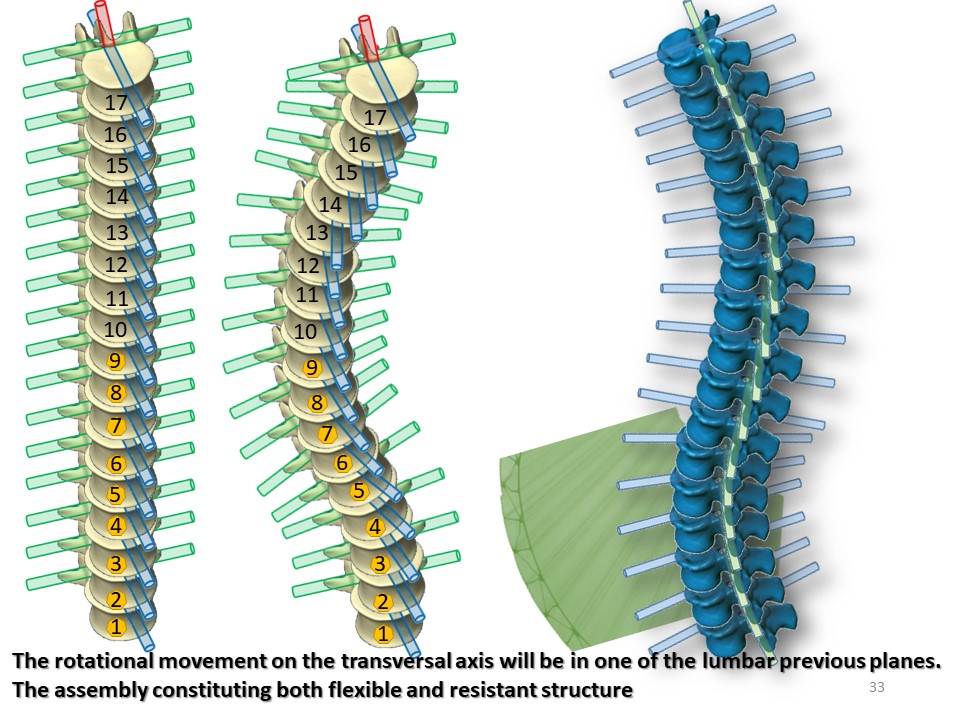 |
In scoliosis, the multiplicity of segmental mobility planes of the sagittal plane means that in planar geometry the mechanical action of braces is often limited to the apical scoliotic vertebra. |
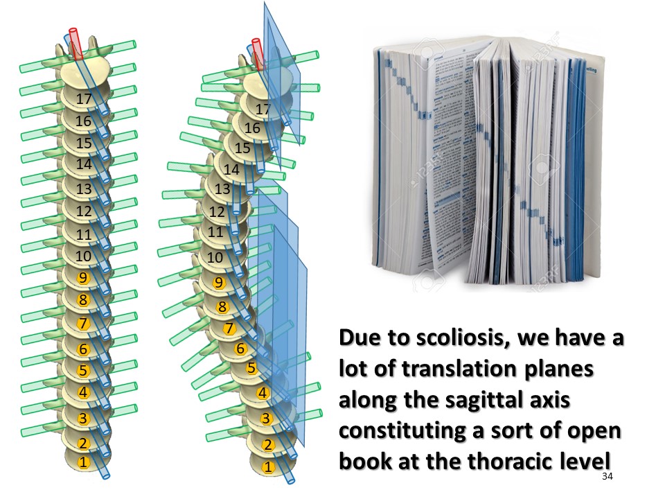 |
It is the same at the thoracic level, the multiplicity of sagittal mobility planes constitutes a kind of open book, which explains the difficulty of kyphotisation with a flat back. |
 |
In the sagittal plane, mobility is facilitated at the lumbar level, more limited at the thoracic level as much as the thickness of the discs is less. |
 |
The correction in the sagittal plane is however fundamental to obtain the isostatic balance, because it is in this ideal contact position that the best mobility in the frontal plane is obtained. |
Comment in the certification part of the website
|
|
 |
|
 |
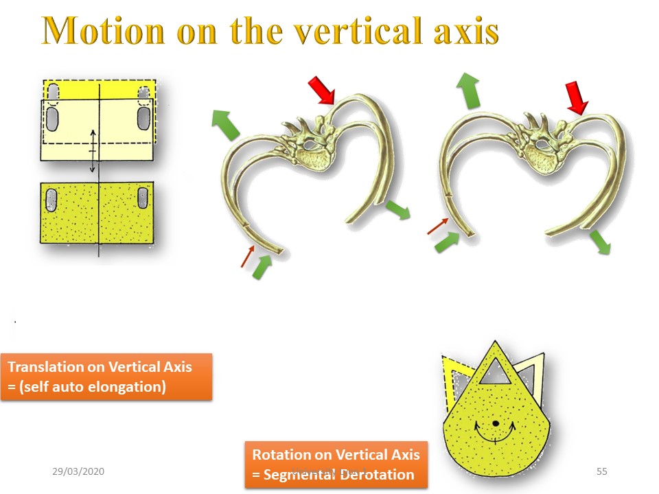
The torsion is the result of the sum of the rotations along the vertical axis. It is favoured by gravity. |
 |
The movement in the horizontal plane is the sum of all intervertebral movements at the lumbar, thoracic and cervical level |
Comment in the certification part of the website
|
|
 |
|
 |
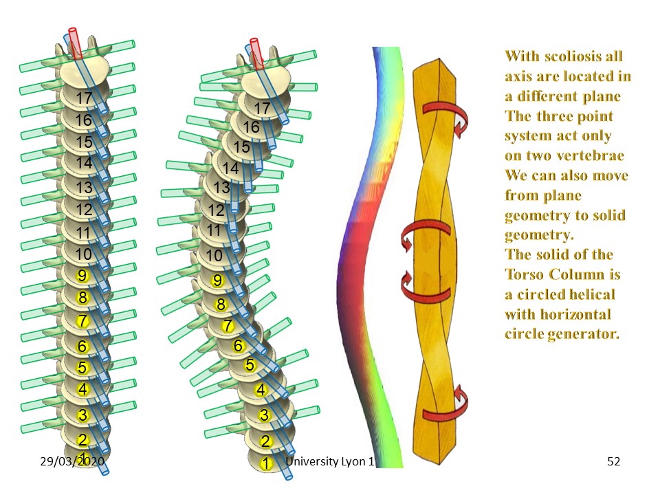
The movement along the vertical axis is characteristic of the torso column, |
 |
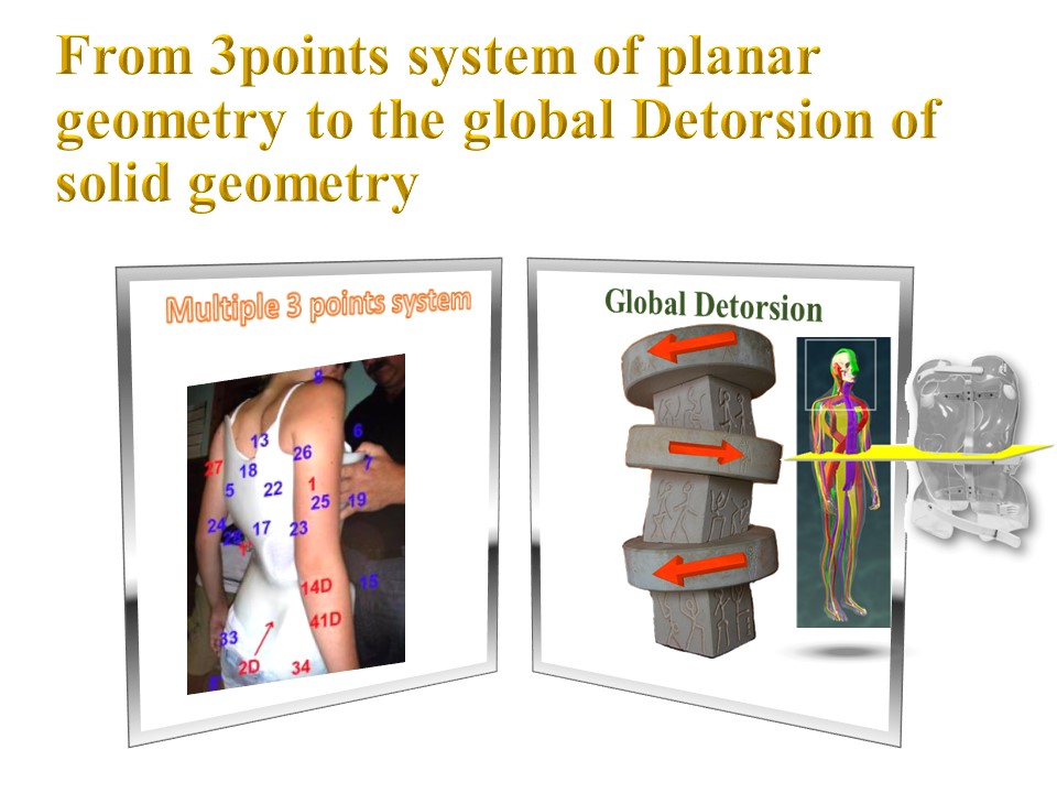
Scoliosis is a helicoid circled with a horizontal generator circle. We will speak of torsion when we consider the whole of a curvature or a vertebral region, we will speak of rotation at a segmental level usually maximum and measured at the level of the apical vertebra. |
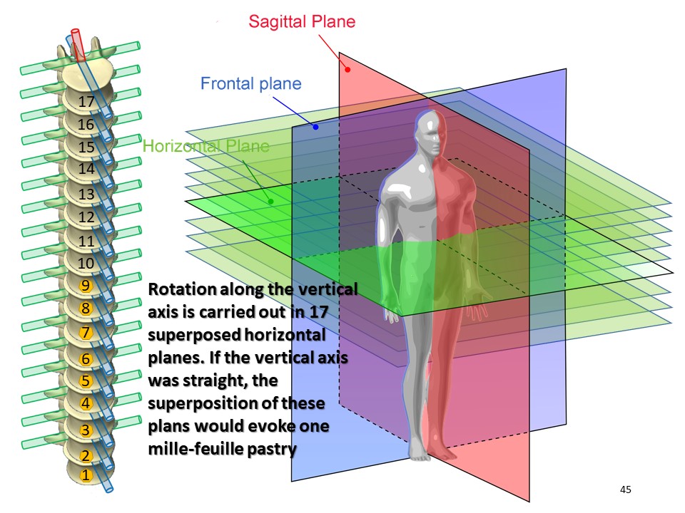 |
The rotation along the vertical axis takes place in 17 superimposed horizontal planes. If the vertical axis were rectilinear, the superposition of these planes would evoke a millefeuille pastryx |
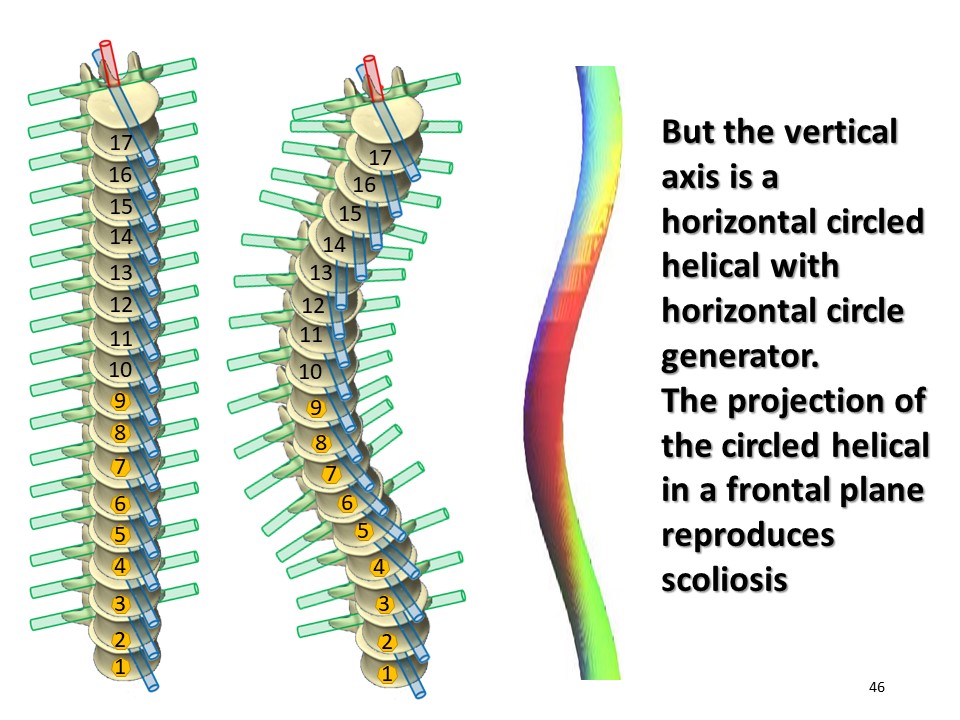 |
In reality in scoliosis, the vertical axis is not rectilinear, and it is impossible to obtain a distortion in plane geometry, |
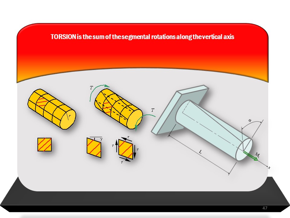 |
The torsion is the sum of the segmental rotations which are all in a different plane. |
 |
|
| > | Comment in the certification part of the website
|
 |
Overall derotation or untwisting is the sum of the translation along the vertical axis (push up effect) and the rotation around the vertical axis induced by asymmetry. |
Comment in the certification part of the website
|
|
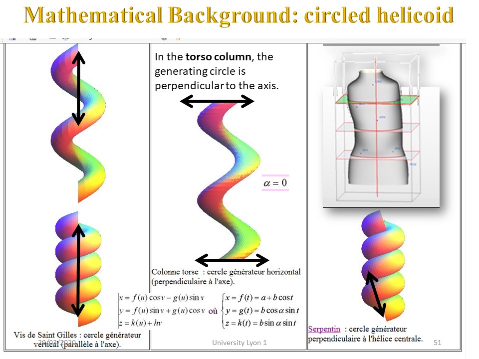 |
The advantage of the helicoid circled with a horizontal generator circle is that it is possible to modify and superimpose several vertebral regions without losing the helical movement. |
 |
The advantage of the helicoid circled with a horizontal generator circle is that it is possible to modify and superimpose several vertebral regions without losing the helical movement. |
 |
The torsion will take place between two fixed points at the two ends of the curvature. |
In solid geometry, there are two types of twist, geometric twist and mechanical twist. |
|
 |
The first untwisting mechanism is geometrical detorsion, that is to say, elongation by translation along the vertical axis in the opposite direction from compression-buckling. |
 |
The maximum space at the level of the intervertebral disc increases mobility and promotes segmental derotation. At the thoracic level, derotation is carried out preferably by the concavity to promote the isostatic balance. |
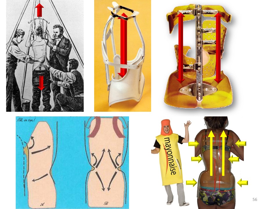 |
Axial elongation has always been an important part of conservative orthopaedic treatment for scoliosis. The fixed point is almost always the pelvis. In the former Lyon Stagnara brace, the axial elongation is obtained by adjustable underarm supports along the rear mast. In Boston and Chêneau, it is the lower part of the spine which favours the extension (cherry stone effect) In ARTbrace, it is the precise application at the sub axillary level and the adjustment of the rack closure at thoraco-lumbar level which causes a kind of extrusion (mayonnaise tube effect),x |
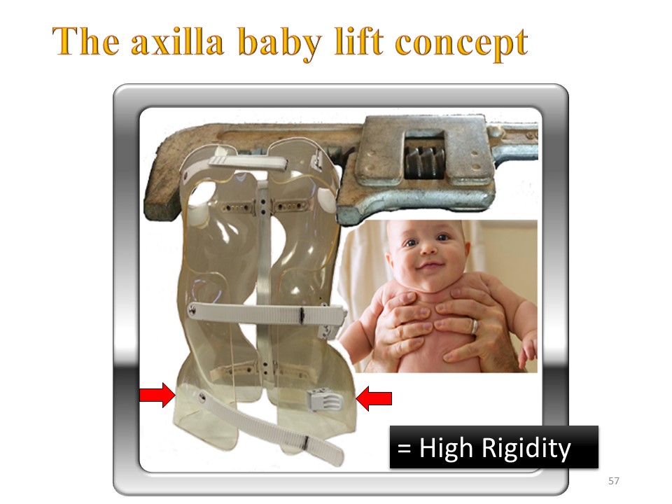 |
In ARTbrace, the under-axillary grip is like to lift a child. The high rigidity of the polyamide alone ensures this grip without the need to use the anterior closure by scratch which is only there to stabilize the brace. |
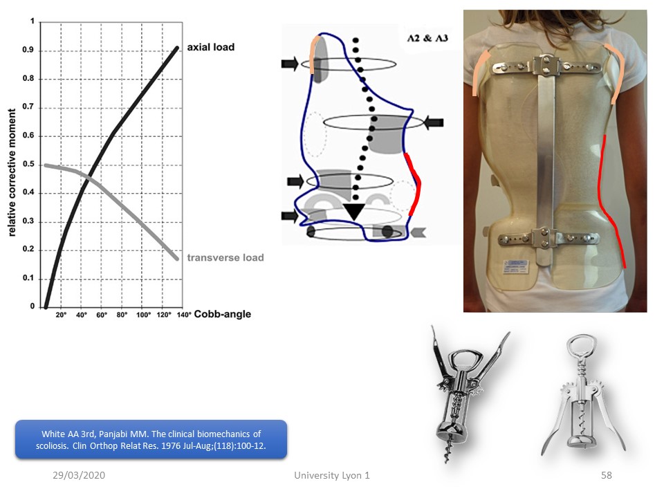 |
This graph by Panjabi and White shows the predominant effectiveness of mechanical actions along the vertical axis beyond 40 °. |
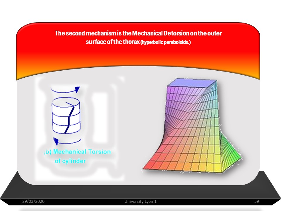 |
The second mechanism is the mechanical detorsion, which takes place across the entire external surface of the rib cage. The brace will correct the chest deformation and the asymmetry of the waist fold, but above all move the volumes. The faces of the solid obtained in the cutting planes are called the bases of the trunk, and the distance between the two cutting planes is the height of the trunk. The lateral faces of this solid are not necessarily planar but are hyperbolic paraboloids. |
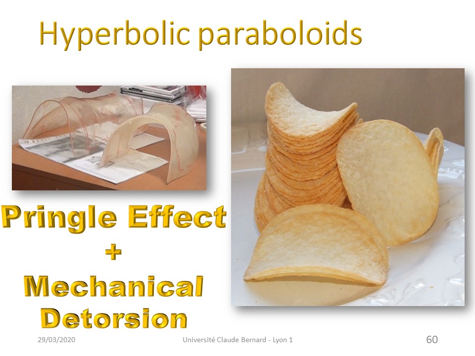 |
The two hemi-pieces of ART are indeed hyperbolic paraboloids adding "Pringle effect" and mechanical detorsion obtained by the detorsion of moldings 2 and 3 |
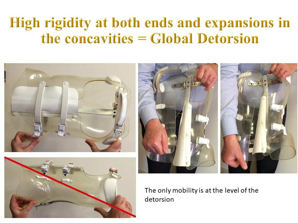 |
In the frontal plane, the only mobility takes place in the expansions. In the sagittal plane, there is no mobility. On the other hand, shearing and derotation are possible depending on the deformation of the metal assembly of the rear bar and hinges and of the polyamide hemi-valves. |
 |
|
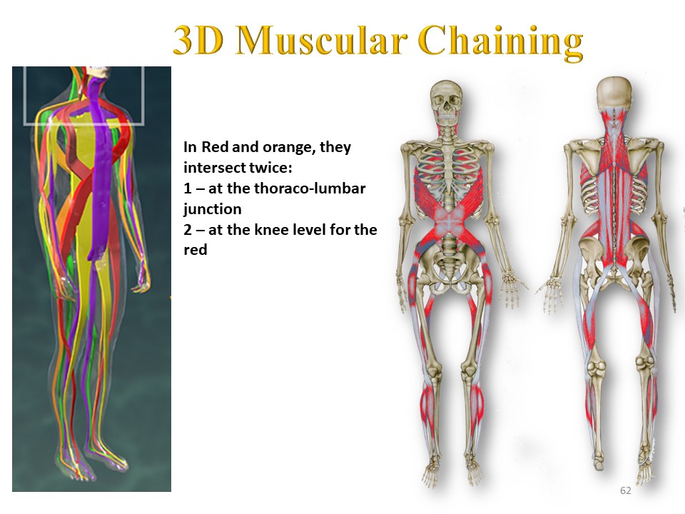 |
Clinically and according to the same principle muscles and fascias constitute spiral chains crossing forward at the thoraco-lumbar level and behind at the lumbo-sacral level. |
Comment in the certification part of the website
|
|
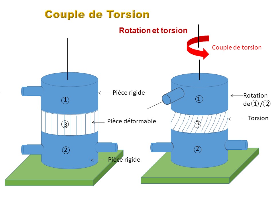 |
Between the two fixed points corresponding to the limits of scoliosis, a couple of detorsion is performed. |
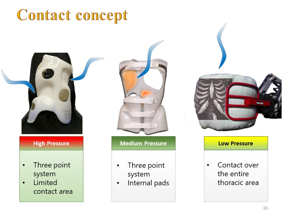 |
The type of contact is a major element of compliance. It can be compared to that achieved for lifting hay bales without tearing the thin plastic wrap that covers them. There is no longer the high-pressure contact created by the multiple 3 points system, but a global untwisting between the two axillary and pelvic wrenches. |
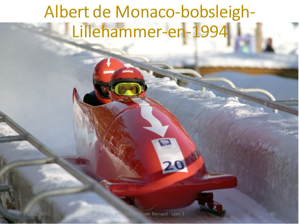 |
In asymmetrical braces with high rigidity, there are no more pads. The trunk is guided and must move as in a bobsleigh track in the opposite direction of scoliosis. |
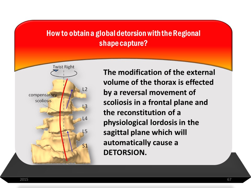 |
The modification of the external volume of the trunk is done by a reversal movement of the scoliosis in a frontal plane and the reconstitution of a physiological sagittal plane in isostatic balance which will automatically cause DETORSION. |
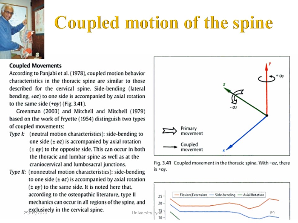 |
This detorsion is the consequence of the coupled movements in the sagittal and frontal plane, as Panjabi explained in 1978. |
 |
|
 |
This distortion will be maximum in sagittal isostatic balance. |
Comment in the certification part of the website
|
|
 |
The laws of coupled movements at the level of the spine have also been described in the context of vertebral manipulations. For the same frontal inflection, the direction of rotation can vary depending on the sagittal plane. To obtain a derotation with scoliosis, the sagittal plane must be positioned in isostatic balance. In case of a flat back, the scoliotic rotation can be accentuated. |
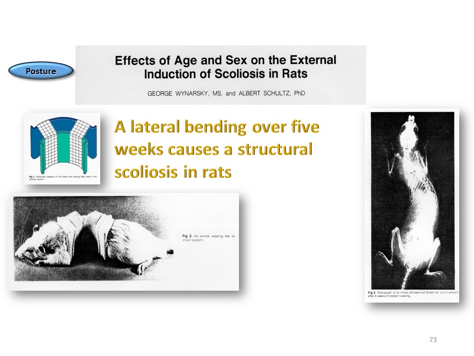 |
The illustration of this principle is obtained experimentally by creating a structural scoliosis with a single bending in the frontal plane. |
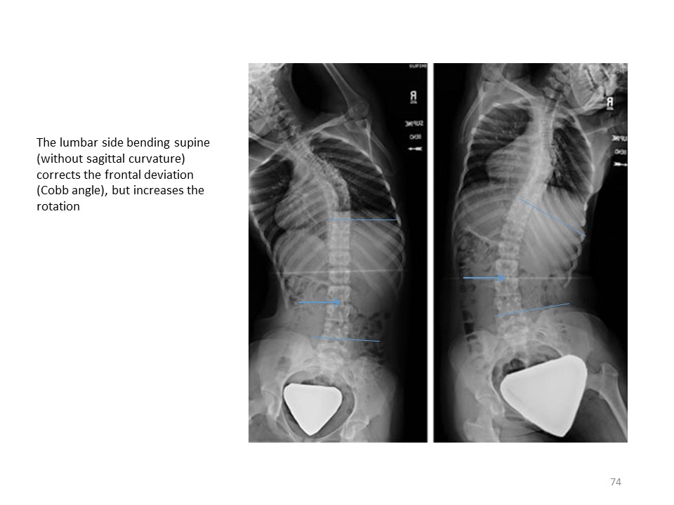 |
In some corrective bendings performed in the supine position, the lumbar rotation can be accentuated. |
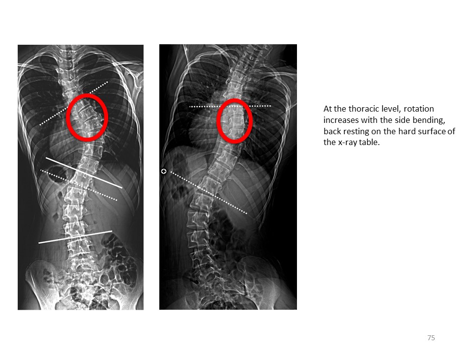 |
It is the same here at the thoracic level. |
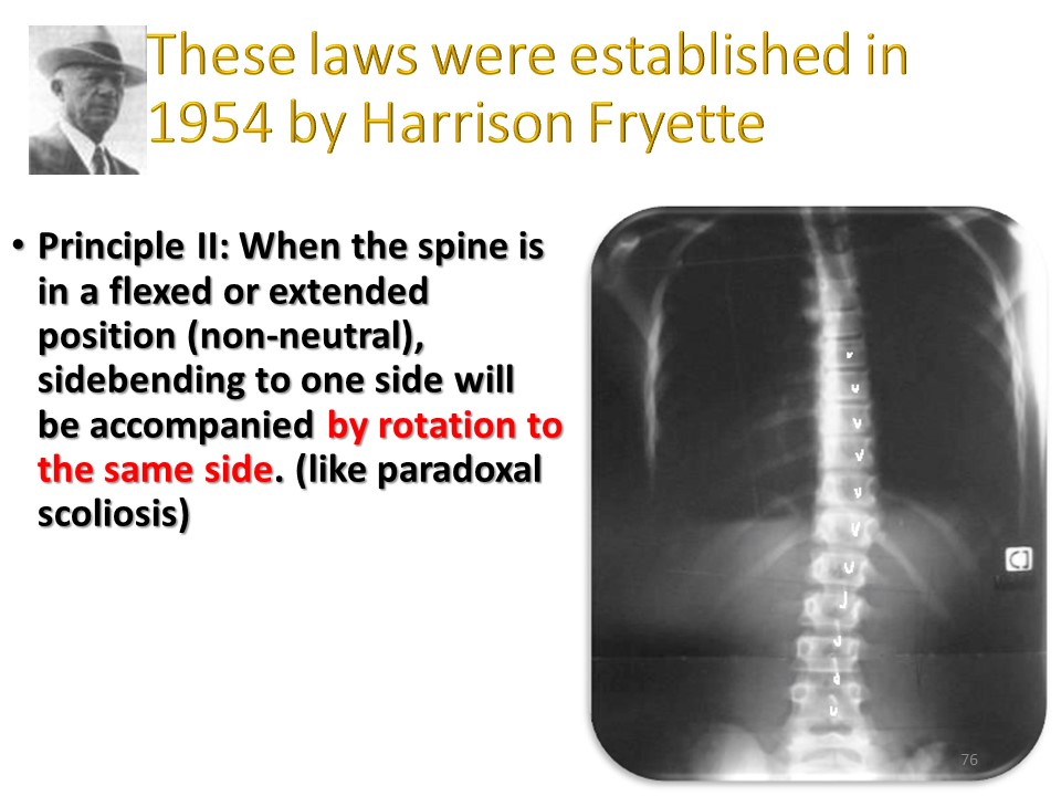 |
According to the 2nd principle of vertebral manipulations, when the spine is curved in flexion or extension in the sagittal plane, that is to say in isostatic balance, a unilateral inflection is accompanied by a rotation in the same direction, correcting the scoliotic rotation. |
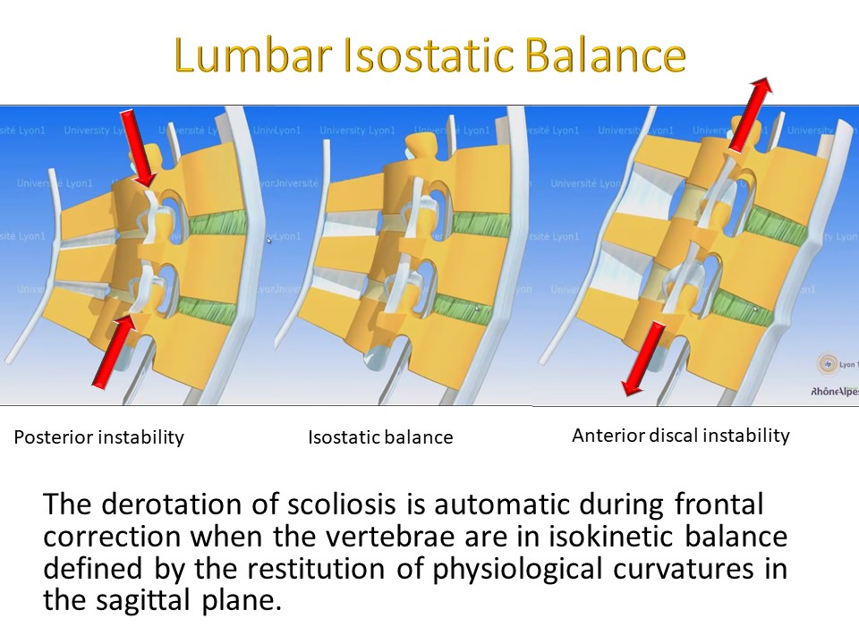 |
The derotation of scoliosis is automatic during frontal correction when the vertebrae are in isostatic balance defined by the restitution of physiological curvatures in the sagittal plane. |
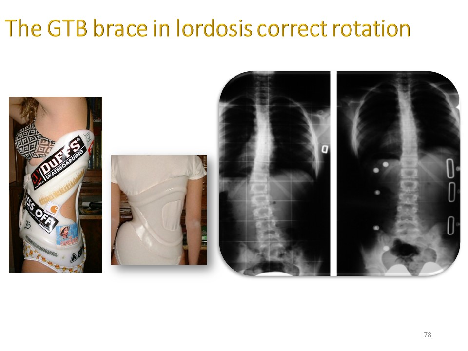 |
The GTB lumbar brace is always made in lordosis and the derotation is clearly visible in this example. |
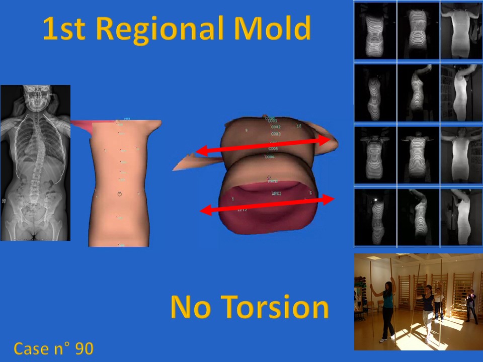 |
Confirmation is provided by studying the 3 successive scans produced for ARTbrace. During the moulding n ° 1 carried out in extension, the two girdles remain balanced without rotation. |
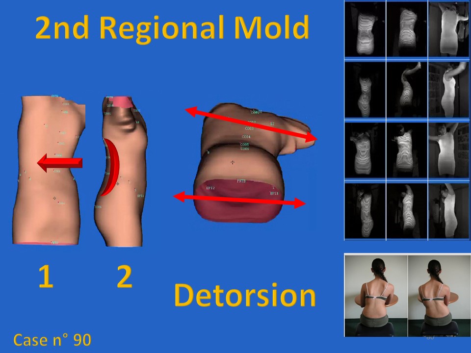 |
During scan No. 2 at the lumbar level, a first detorsion is noted between the pelvic girdle and the shoulder girdle. |
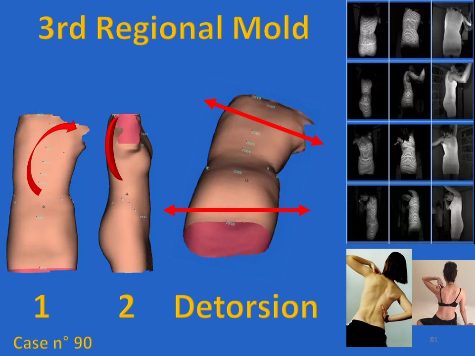 |
This detorsion between the two girdles is accentuated during scan No. 3 performed at the thoracic level. |
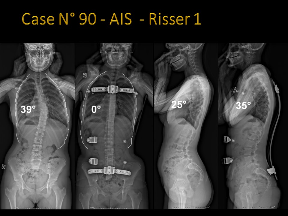 |
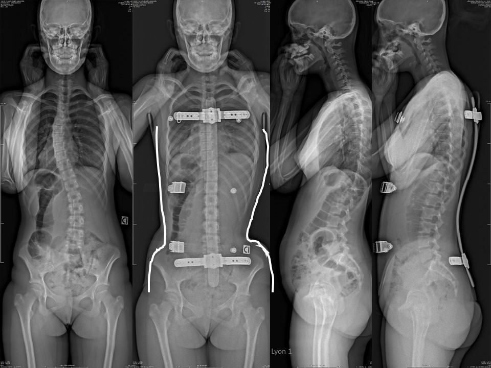
Here is the result of this case n° 90, with initially thoracic scoliosis of 39° and rib hump of 17° ATR. Excellent in-brace correction with total reduction of the curvature. The sagittal plane is also improved, |
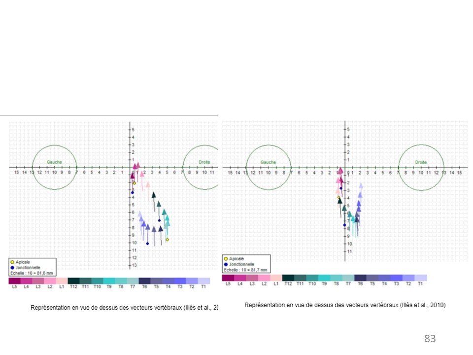 |
Under EOS 3D, detorsion is excellent with centring of the vertebrae on the midline |
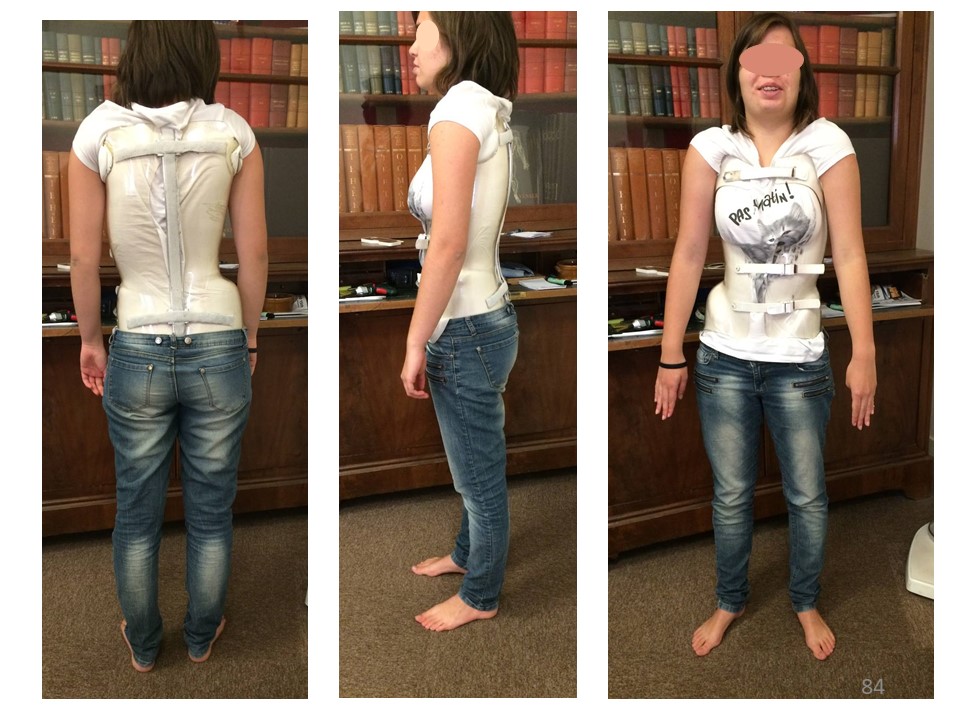 |
Here is the Clinical picture in brace. |
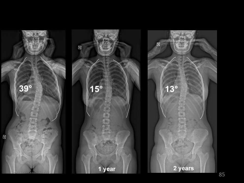 |
At Risser 5, after 2 years of bracing, the angulation is 13°, |
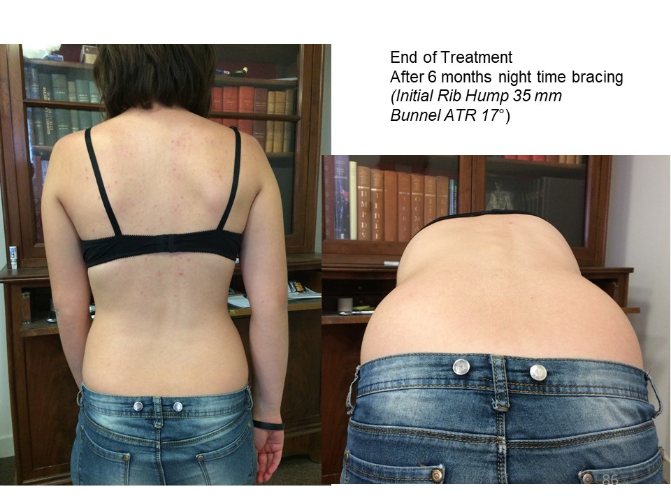 |
Clinical aspect at the end of treatment after 6 months of nighttime bracing The Bunnel ATR is under 10°. |
 |
The most dramatic example was noted in this 11-year-old child living in Brazil who has a large thoracic rotation. The in-brace correction is satisfactory. |
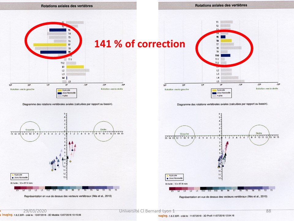 |
But the most spectacular correction lies in the horizontal plane with inversion of segmental rotations and global torsion. As always, the vertebrae cluster along the line of gravity. |
 |
In conclusion, the Lyon method follows directly from the 3D biomechanics of scoliosis and shows the interest of the global geometry of solids with regional 3D correction by coupled movements. ARTbrace physiotherapy and corset follow exactly the same principles. |
 |